When you bring a dog into your home, understanding their lifelong health trajectory helps you shield them from preventable suffering.
Many popular canine breeds carry inherited genetic mutations that put them at heightened risk for serious ocular conditions, ranging from progressive vision loss to chronic surface inflammation.
Identifying these hereditary vulnerabilities early allows you to schedule targeted veterinary screenings, implement preventive eye care routines, and select responsible breeders who perform certified DNA testing.
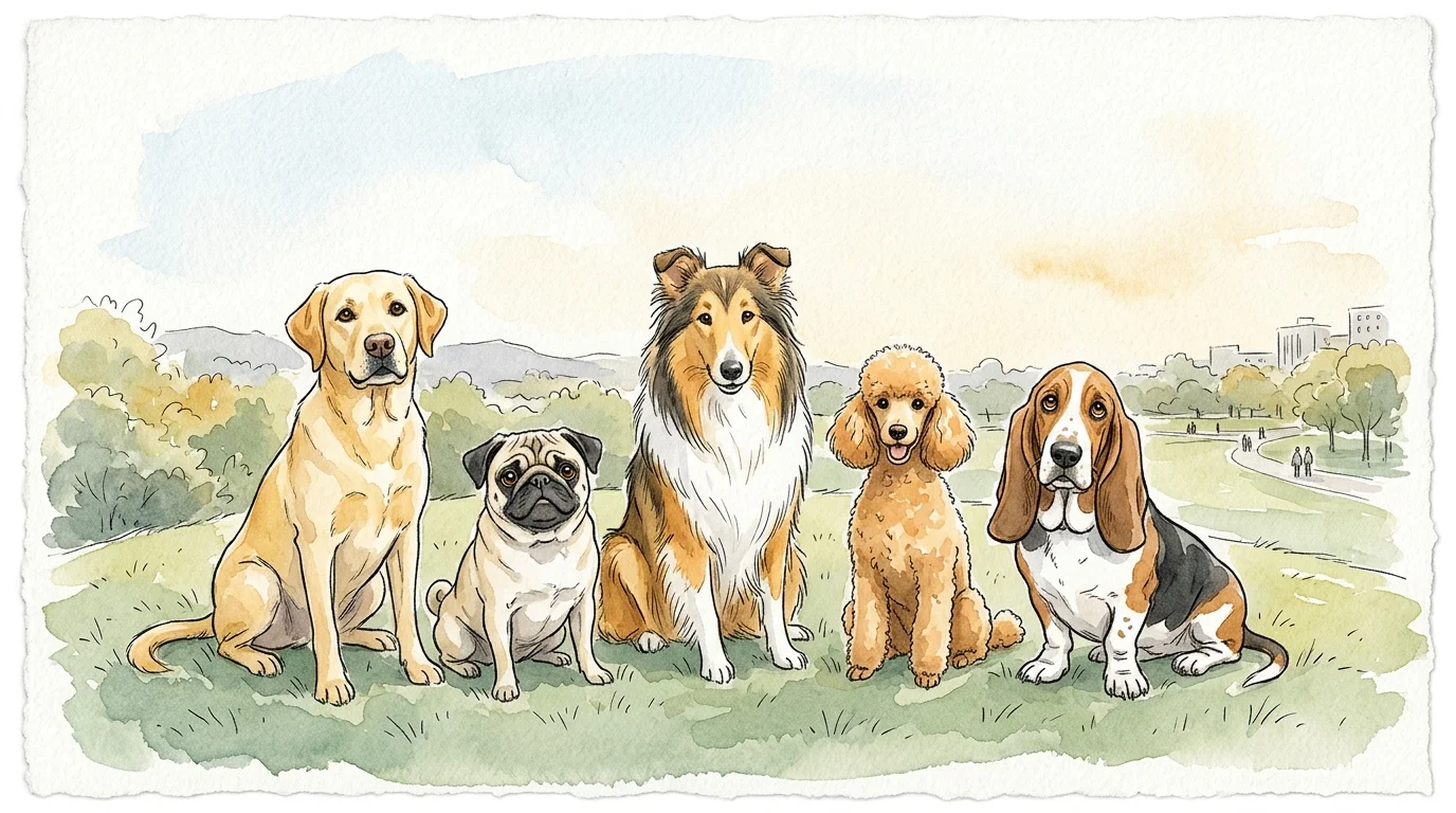
Whether you care for an energetic retriever or a flat-faced companion, learning about dog breeds prone to eye problems empowers you to safeguard your dog’s sight for years to come.
Understanding Hereditary Eye Diseases in Dogs
Hereditary eye disease in dogs stems from genetic variations that passed down through generations of purebred breeding lines.
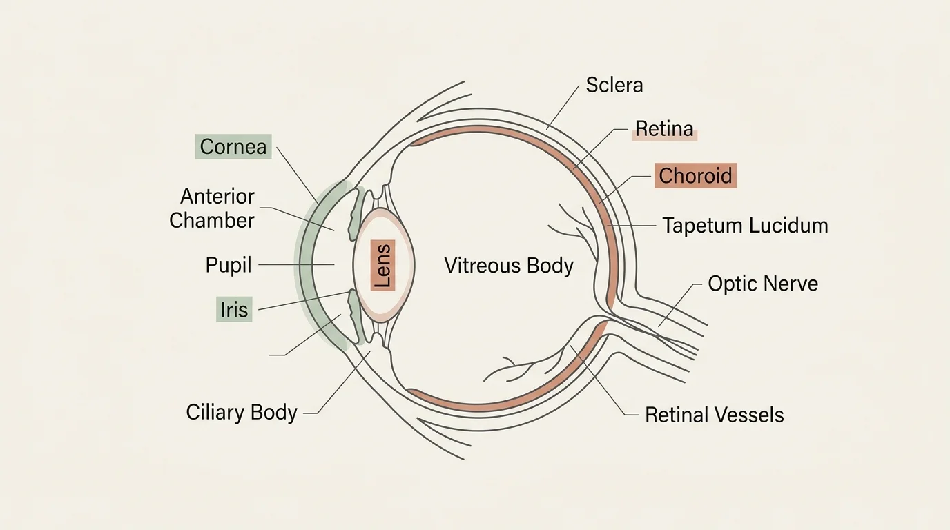
These mutations affect different anatomical structures within the eye; some damage the light-sensing retina at the back of the globe, while others impair the cornea, clear lens, or eyelid alignment at the front.
Genetic conditions generally fall into two categories: developmental structural defects and progressive degenerative diseases.
Developmental defects exist at birth or manifest during early puppyhood. Conditions like Collie Eye Anomaly (CEA) or hereditary juvenile cataracts occur when specific genes alter normal ocular tissue formation.
Progressive degenerative diseases, such as Progressive Retinal Atrophy (PRA), gradually destroy specialized rod and cone cells over time; affected dogs often appear healthy for several years before showing clinical night blindness.
Conformation plays a substantial role alongside direct gene mutations. Brachycephalic (short-muzzled) breeds feature shallow eye sockets that expose a large percentage of the globe to air, dust, and mechanical friction.
According to the American College of Veterinary Ophthalmologists (ACVO), combining structural facial traits with inherited tear-film deficiencies creates a compound risk for chronic corneal damage and vision loss.
01 Rough Collie: Collie Eye Anomaly (CEA)
The Rough Collie remains the flagship breed associated with Collie Eye Anomaly (CEA), an autosomal recessive disorder caused by a mutation in the NHEJ1 gene. This mutation disrupts normal development of the choroid—the vital layer of blood vessels that nourishes the retina.
Genetic data collected from 2010 through 2023 indicates that between 75% and 90% of Rough Collies in the United States carry at least one copy of this gene mutation or express the clinical disease.
CEA manifests along a wide spectrum of severity, even among littermates carrying the same genetic variation:
- Choroidal Hypoplasia: The mildest form, where underdeveloped blood vessels create thin patches in the tissue; this often causes minimal visual disruption.
- Coloboma: A pit or defect in the optic nerve or surrounding tissue that can predispose the retina to detachment.
- Retinal Detachment: Severe structural defects cause the retina to separate, leading to complete and sudden blindness in the affected eye.
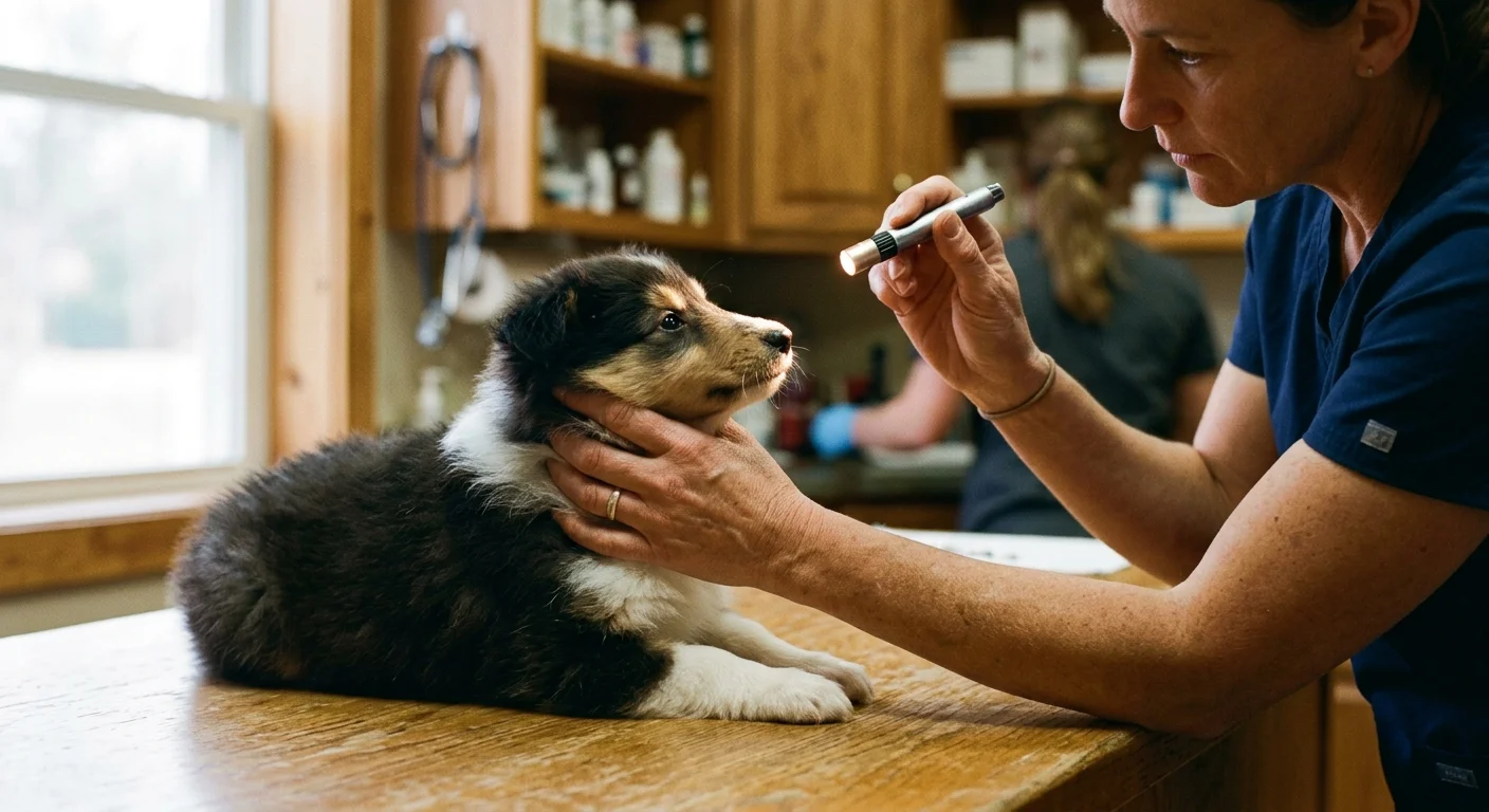
Because choroidal pigment develops as a puppy matures, veterinary ophthalmologists must screen Collie litters between 5 and 8 weeks of age. Early screening catches visible tissue defects before pigment hides them.
While CEA has no cure, DNA testing allows breeders to test breeding pairs and prevent passing two copies of the mutant gene to offspring.
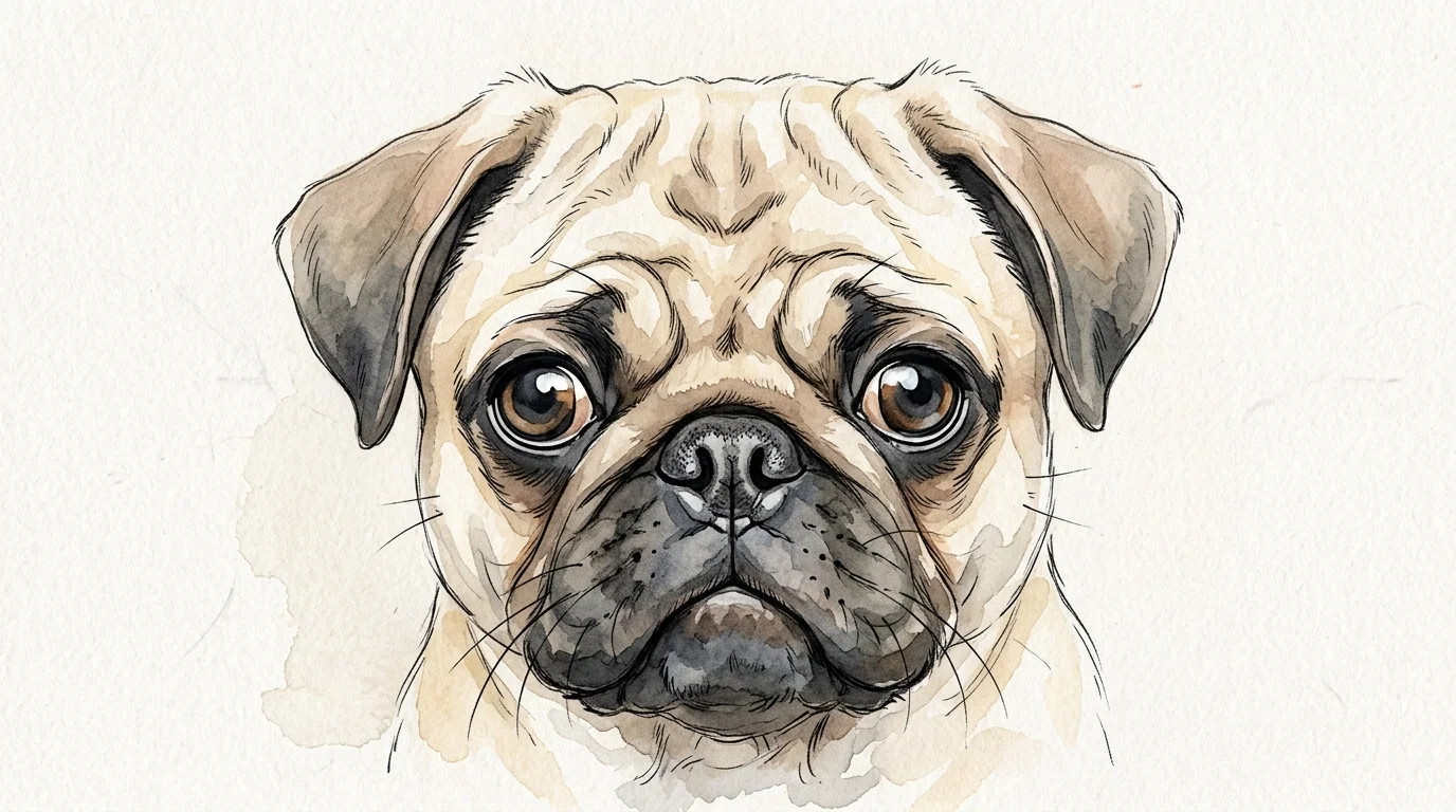
02 Pug: Pigmentary Keratopathy and Brachycephalic Ocular Syndrome
Pugs possess prominent, appealing eyes, but their brachycephalic facial structure introduces severe ocular surface risks. Shallow eye sockets result in exophthalmos (bulging eyes), preventing complete eyelid closure when the dog blinks or sleeps.
This constant exposure causes chronic dryness, inflammation, and repeated micro-trauma from eyelashes and facial skin folds.
A study published in the Journal of Veterinary Internal Medicine evaluating 295 Pugs revealed that 82.4% had corneal pigmentation (pigmentary keratopathy) in at least one eye. The body deposits brown melanin pigment across the cornea in response to ongoing irritation.
Over months and years, this pigment sheets across the clear tissue, eventually blocking light from entering the eye entirely.
Data from a 2025 VetCompass study analyzing 1,318 Pugs found that 14.5% suffered from eye or eyelid disorders. The most widespread conditions were corneal ulcers (5.5%), corneal pigmentation (3.6%), and Keratoconjunctivitis Sicca, commonly known as dry eye (3.3%).
You can protect a Pug’s sight by applying daily lubricating eye drops prescribed by your veterinarian and seeking immediate medical care if your dog squints or rubs their face.
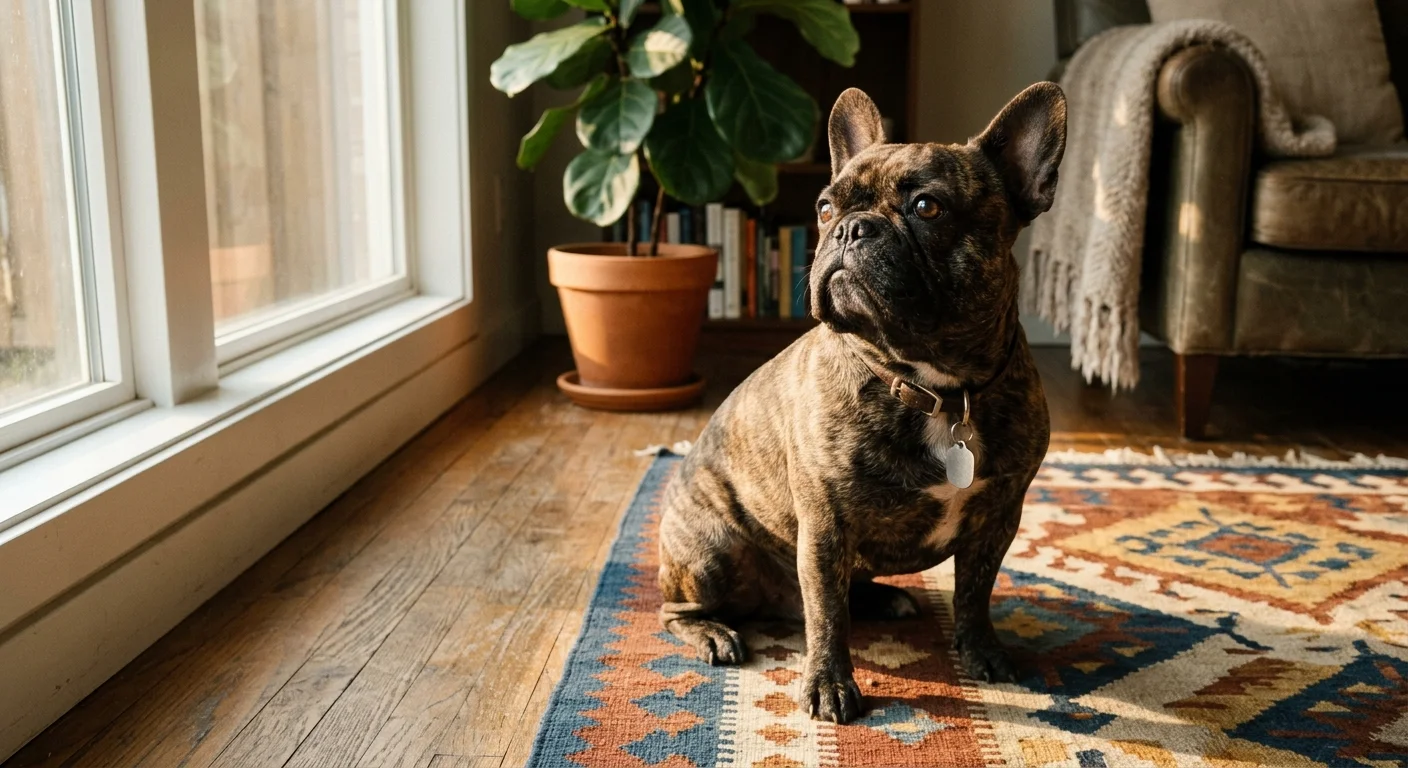
03 French Bulldog: Ocular Surface Disorders and Keratitis
French Bulldogs share similar anatomical risks with Pugs, but clinical data demonstrates an exceptionally high rate of surface inflammation and tear film instability. Their wide eye openings and facial wrinkles create an environment where dust and bacteria trigger persistent inflammatory responses.
A clinical study examining 809 French Bulldogs revealed that 76.6% presented with keratitis (corneal inflammation or ulceration) and 45% suffered from tear film disorders like dry eye.
When a dog produces inadequate quality or quantity of tears, the delicate corneal epithelium becomes dry, cloudy, and prone to painful ulcerations that can erode deep into the cornea within 24 to 48 hours.
French Bulldogs also suffer from cherry eye—a prolapse of the third eyelid gland—and inherited juvenile cataracts. You should routinely inspect your French Bulldog’s eyes for redness, discharge, or surface cloudiness.
Regular tear production testing (Schirmer Tear Test) during routine veterinary visits helps catch dry eye before chronic keratitis permanently scars the corneal surface.
04 Labrador Retriever: Progressive Rod-Cone Degeneration (PRCD-PRA)
Labrador Retrievers are widely celebrated for their working intelligence and family temperament, but hereditary eye disease dogs in this category frequently carry genes for Progressive Retinal Atrophy (PRA). PRA represents a broad family of inherited diseases that destroy the photoreceptor cells in the retina.
In Labradors, the primary form is Progressive Rod-Cone Degeneration (PRCD-PRA).
PRA is an incurable, painless disorder affecting over 100 dog breeds across more than 60 identified gene mutations. In PRCD-PRA, rod cells—which detect motion and govern night vision—die first.
Affected Labradors gradually lose the ability to navigate outside at night or see in dimly lit rooms. Over the following months or years, cone cells degenerate as well, leading to total blindness.
A 10-year genetic study published in 2023 showed that over 15% of tested Labrador Retrievers were heterozygous carriers for PRCD-PRA. Carriers do not develop vision loss themselves, but passing the gene to offspring can result in blind puppies if bred to another carrier.
DNA panel testing allows responsible breeders to eliminate carrier-to-carrier pairings entirely.
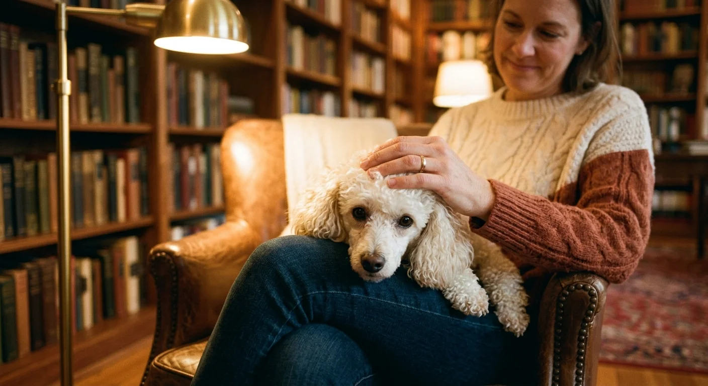
05 Toy and Miniature Poodle: PRCD-PRA and Hereditary Cataracts
Toy and Miniature Poodles feature prominently on the list of dog breeds prone to eye problems due to a high incidence of PRCD-PRA and inherited cataracts.
Like Labradors, the 2023 genetic study confirmed that over 15% of tested Toy Poodles carry the mutant PRCD gene responsible for adult-onset retinal degeneration.
In addition to PRA, Poodles carry genetic predispositions for early-onset hereditary cataracts. A cataract is an opacity within the eye’s crystalline lens that obstructs light transmission to the retina.
In Poodles, genetic cataracts can develop as early as 1 to 3 years of age, rapidly progressing from small specks to total lens cloudiness.
Unlike PRA, cataracts can often be treated surgically through phacoemulsification—a procedure where a veterinary ophthalmologist breaks down the cloudy lens with ultrasound waves and implants an artificial intraocular lens.
Routine eye exams help identify early cataracts before they cause secondary lens-induced uveitis (severe internal inflammation).
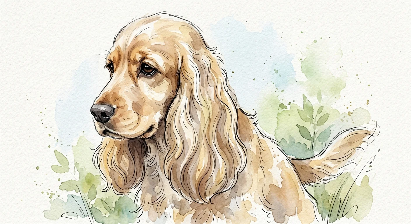
06 Cocker Spaniel: Primary Glaucoma and Distichiasis
Both American and English Cocker Spaniels face inherited risks for primary glaucoma, a severe condition characterized by improper drainage of fluid within the eye. In healthy eyes, aqueous humor flows continuously to maintain eye shape and nourish internal structures.
In dogs with primary glaucoma, abnormal development of the iridocorneal angle restricts fluid outflow, causing intraocular pressure (IOP) to spike rapidly.
Elevated intraocular pressure causes intense pain and destroys the sensitive optic nerve fibers within hours. Cocker Spaniels often present with acute primary closed-angle glaucoma, where pressure surges suddenly, turning the eye red, cloudy, and unresponsive to light.
Emergency veterinary treatment is critical to preserve sight when a pressure spike occurs.
Cocker Spaniels also suffer from distichiasis, an inherited condition where extra eyelashes grow from the Meibomian glands along the eyelid margin. These misdirected hairs rub continuously against the cornea, causing tearing, squinting, and corneal ulcers if left unmanaged by a vet.
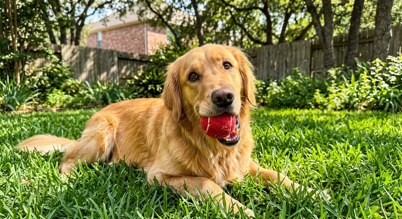
07 Golden Retriever: Pigmentary Uveitis
Golden Retrievers possess a unique, breed-specific hereditary eye disease known as Golden Retriever Pigmentary Uveitis (GRPU). GRPU is a late-onset inflammatory condition that typically affects middle-aged and senior Golden Retrievers, usually appearing around 8 to 10 years of age.
GRPU manifests through distinct clinical signs that develop subtly inside the eye:
- Fluid-Filled Cysts: Tiny, translucent uveal cysts float within the anterior chamber or hide behind the iris.
- Spoke-like Pigment: Melanin pigment deposits in a radial, spoke-like pattern on the front surface of the lens.
- Secondary Glaucoma: Inflammatory debris blocks the eye’s fluid drainage pathways, driving up intraocular pressure.
Because GRPU develops late in life, dogs often pass the gene to multiple generations before showing symptoms. Annual screening by a board-certified veterinary ophthalmologist starting at age 3 ensures early detection.
Prophylactic anti-inflammatory eye drops can slow disease progression and delay secondary glaucoma.
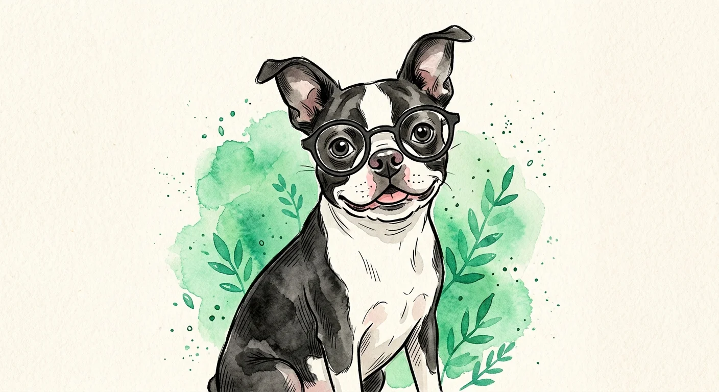
08 Boston Terrier: Early-Onset Juvenile Cataracts
Boston Terriers carry genetic eye risks stemming from both their brachycephalic head structure and explicit gene mutations. The breed is particularly known for autosomal recessive juvenile cataracts caused by a mutation in the HSF4 gene.
Affected puppies develop dense cataracts in both eyes as early as 3 to 12 months of age, leading to functional blindness before adulthood.
Commercial DNA testing for the HSF4 mutation gives breeders a clear method to screen parents before mating. Dogs carrying two copies of the mutation will develop early cataracts, while dogs with one copy remain clear but can pass the mutation down.
Beyond cataracts, Boston Terriers feature large, prominent eyes that face constant risk from mechanical injuries, dry eye, and corneal deep ulcers. You should monitor your Boston Terrier closely when they play outdoors and seek prompt care if you notice cloudiness or rubbing behavior.
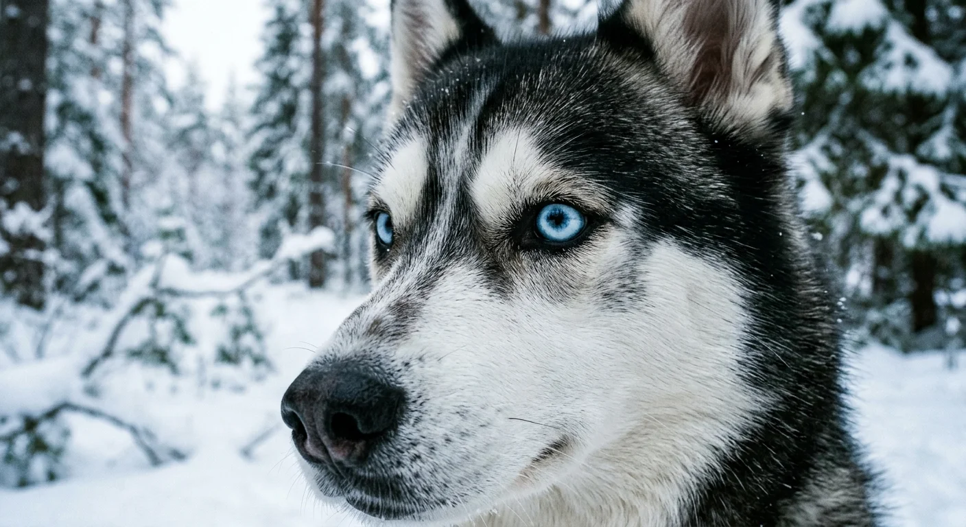
09 Siberian Husky: Hereditary Cataracts and X-Linked PRA
Siberian Huskies are susceptible to hereditary cataracts that differ structurally from those seen in Poodles or Boston Terriers. Husky cataracts usually develop in the posterior cortex of the lens between 6 and 18 months of age.
While some of these cataracts remain small and do not impair vision, others progress rapidly to blind the dog.
Huskies also carry a unique form of Progressive Retinal Atrophy called X-Linked PRA (XL-PRA). Because the defective gene resides on the X chromosome, male Huskies (who possess only one X chromosome) develop full retinal degeneration if they inherit the gene from their mothers.
Female Huskies with one mutant X chromosome typically act as asymptomatic carriers or display very mild, localized retinal changes.
Male Huskies affected by XL-PRA typically show night blindness around 2 to 4 years of age, progressing to full blindness by age 5. Genetic testing of breeding females is essential to prevent passing this X-linked trait to male offspring.
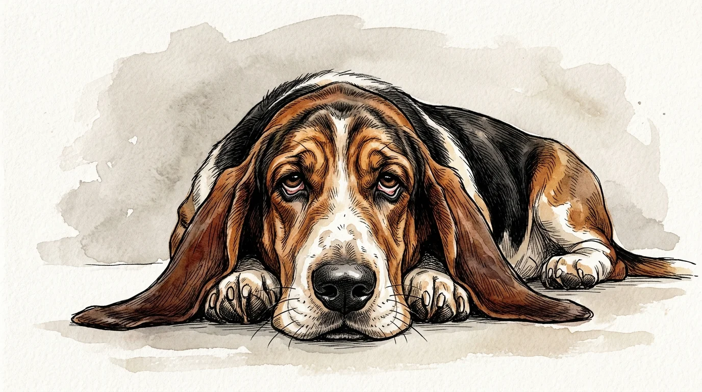
10 Basset Hound: Primary Glaucoma and Eyelid Deformities
The Basset Hound’s iconic loose, heavy facial skin and deep ocular sockets make them vulnerable to multiple severe eye disorders.
Basset Hounds are genetically predisposed to primary open-angle glaucoma and pectinate ligament dysplasia—a structural defect in the drainage angle of the eye that hinders normal aqueous humor outflow.
The breed’s facial structure creates structural eyelid deformities that compound their health risks:
- Ectropion: The lower eyelid sags outward, forming a deep pocket that collects dust, pollen, and bacteria.
- Entropion: The lower or upper eyelid rolls inward, driving eyelashes directly against the sensitive cornea.
Chronic irritation from ectropion and entropion produces pain, mucoid discharge, and corneal scarring. Veterinary surgeons frequently perform corrective eyelid surgery (blepharoplasty) to align the eyelids properly, protecting the cornea from ongoing mechanical trauma and secondary infection.
Screening and Genetic Testing Procedures
Protecting dogs from hereditary eye disease relies on systematic screening protocols established by national veterinary organizations. The Orthopedic Foundation for Animals (OFA) maintains the Companion Animal Eye Registry (CAER), which was established in 2012 to track inherited eye disease trends across dog breeds.
CAER exams must be conducted by Diplomates of the ACVO, who publish updated statistics annually in the ACVO Blue Book.
During a CAER examination, the veterinary specialist dilates the dog’s pupils to inspect the cornea, lens, vitreous, optic nerve, and retina using specialized equipment like slit-lamp biomicroscopes and indirect ophthalmoscopes.
Because many genetic eye diseases appear later in life, dogs used for breeding require annual exams rather than a single lifelong clearance.
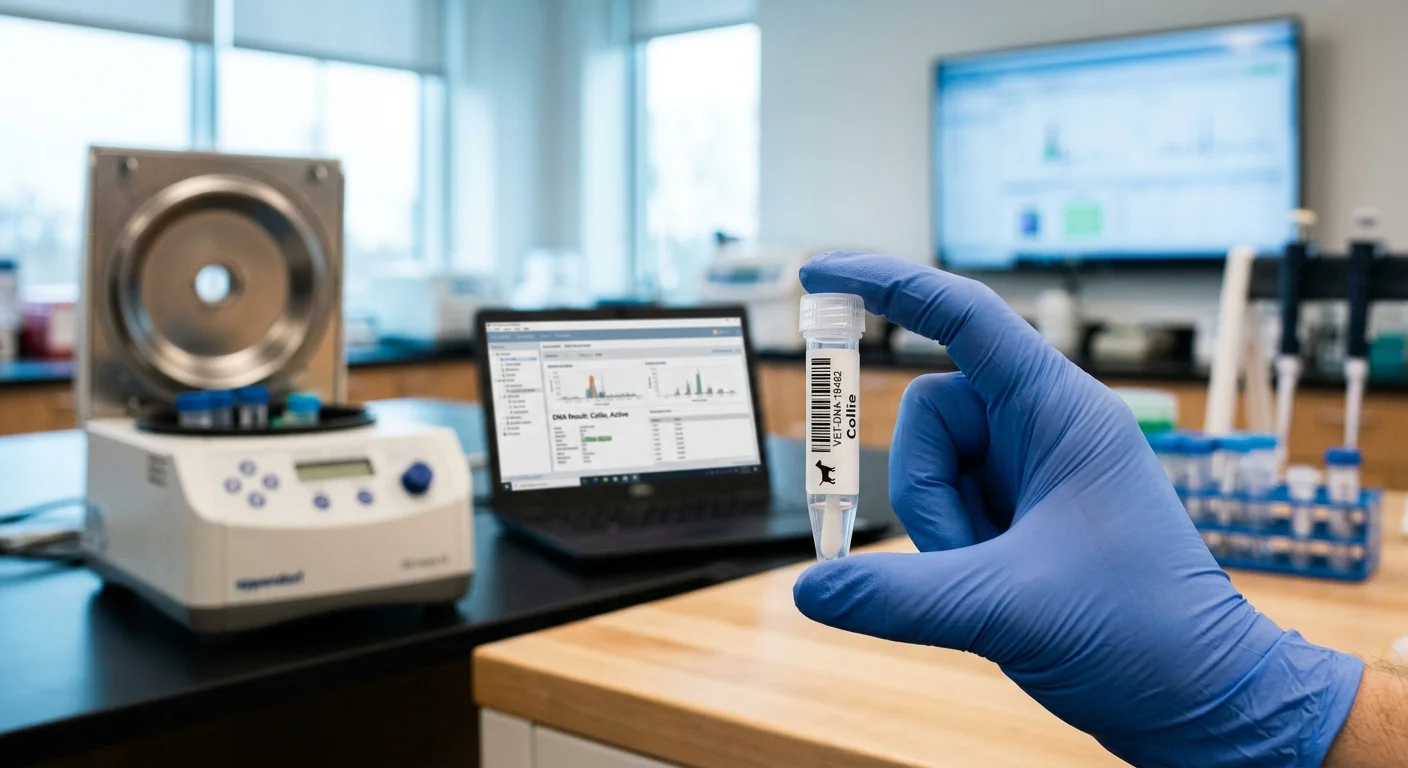
DNA testing complements clinical screening by identifying silent genetic carriers. Modern canine genomic panels detect specific mutations for PRCD-PRA, CEA, and HSF4 juvenile cataracts.
By pairing annual CAER examinations with DNA panel testing, responsible breeders can make informed mating decisions that preserve overall health while working to eradicate inherited blindness.
Comparison of Breed Eye Risks
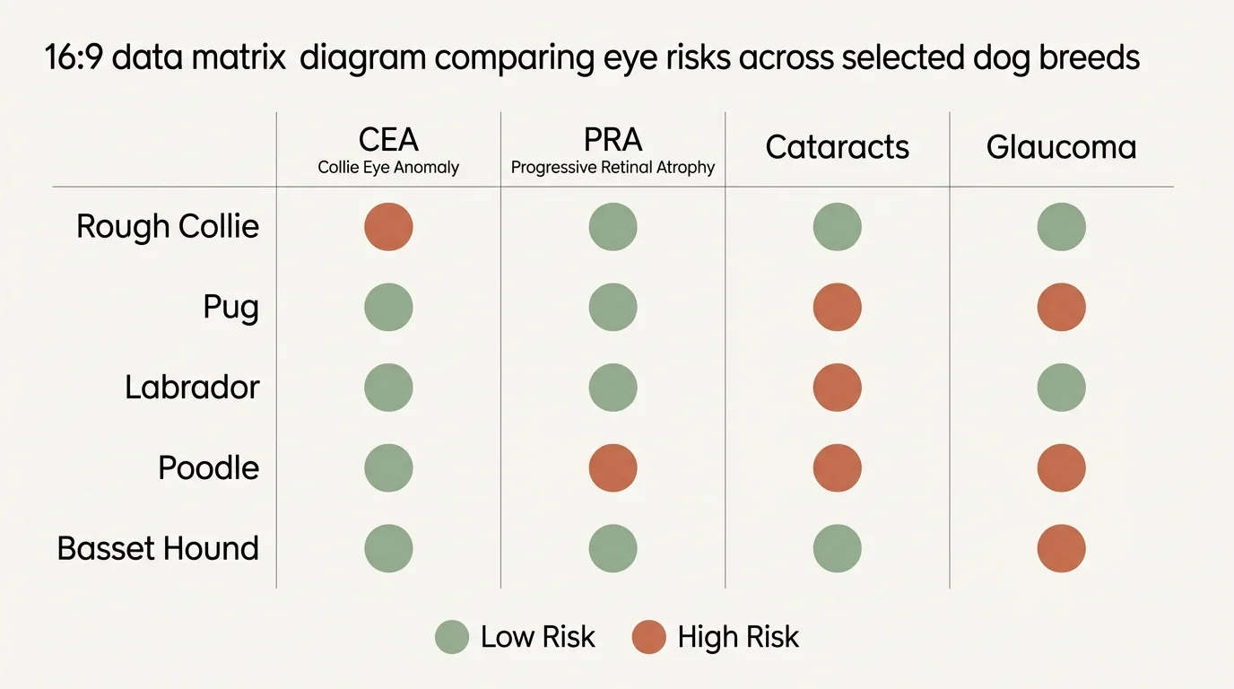
Understanding which risks affect specific breeds helps you choose appropriate diagnostic screenings and daily care strategies for your pet.
| Dog Breed | Primary Genetic Eye Condition | Anatomical / Genetic Root Cause | Recommended Screening Method |
|---|---|---|---|
| Rough Collie | Collie Eye Anomaly (CEA) | Autosomal recessive NHEJ1 gene mutation | Puppy CAER exam (5–8 weeks) & DNA test |
| Pug | Pigmentary Keratopathy | Brachycephalic exposure & lagophthalmos | Annual clinical exam & tear testing |
| French Bulldog | Keratitis & Dry Eye | Shallow orbits & tear film deficiency | Schirmer Tear Test & fluorescein stain |
| Labrador Retriever | PRCD-Progressive Retinal Atrophy | Autosomal recessive photoreceptor decay | DNA testing & annual CAER exam |
| Toy Poodle | PRCD-PRA & Juvenile Cataracts | Genetic mutations affecting retina & lens | DNA panel screening & slit-lamp exam |
| Cocker Spaniel | Primary Glaucoma & Distichiasis | Drainage angle dysplasia & extra lashes | Tonometry (IOP check) & gonioscopy |
| Golden Retriever | Pigmentary Uveitis (GRPU) | Late-onset hereditary uveal cysts | Annual ACVO exam starting at age 3 |
| Boston Terrier | Juvenile Cataracts | Autosomal recessive HSF4 mutation | DNA test for HSF4 mutation |
| Siberian Husky | X-Linked PRA & Cataracts | X-chromosome mutation & cortical opacity | Genetic carrier test for females & CAER exam |
| Basset Hound | Primary Glaucoma & Entropion | Structural drainage defects & loose skin | Annual IOP screening & eyelid evaluation |
What Can Go Wrong: Pitfalls in Canine Eye Health
Navigating hereditary eye health requires vigilance and clear communication with veterinary professionals. Dog owners frequently encounter specific pitfalls that compromise vision:
1. Assuming clear parentage without DNA testing: Purchasing a puppy from parents who look healthy does not guarantee freedom from disease. Recessive conditions like PRA and CEA hide in carrier dogs for generations until two carriers mate.
2. Mistaking early glaucoma for minor eye irritation: Early glaucoma symptoms—such as a slightly red eye, mild cloudiness, or a dog pawing at their face—mimic mild conjunctivitis.
Waiting several days to seek medical attention often results in permanent blindness because elevated intraocular pressure destroys nerve tissue within 24 to 48 hours.
3. Ignoring corneal pigment in flat-faced dogs: Many Pug and French Bulldog owners treat dark corneal spots as normal cosmetic changes. Unmanaged pigmentary keratopathy gradually covers the pupil, causing complete, preventable visual impairment.
4. Administering leftover human or pet eye drops: Applying human eye medications or leftover veterinary steroid drops to a squinting dog can cause devastating harm.
If a dog has an undiagnosed corneal ulcer, topical steroids prevent healing and cause the cornea to dissolve, leading to eye rupture.

Where Outside Advice Pays Off
General practice veterinarians handle many routine ocular problems, but complex hereditary diseases require specialized ophthalmic expertise. Consulting a board-certified veterinary ophthalmologist is essential in specific scenarios:
1. Unexplained vision changes or opacity: If your young or middle-aged dog hesitates in dim light, bumps into doorframes, or develops sudden cloudiness, an ophthalmologist can use specialized equipment to pinpoint whether the issue stems from cataracts, PRA, or corneal dystrophy.
2. Managing chronic, high-risk conditions: Conditions like Golden Retriever Pigmentary Uveitis or primary glaucoma require ongoing intraocular pressure monitoring, advanced medical management, or surgical intervention to maintain comfort and sight.
3. Evaluating breeding dogs: Breeders seeking to clear stock must obtain official CAER examinations from a certified specialist to ensure hereditary conditions are not passed to future litters.
4. Specialized surgical procedures: Cataract removal via phacoemulsification, laser goniotomy for glaucoma, and complex eyelid reconstruction require expert surgical skills and specialized microsurgical tools.
Frequently Asked Questions
Can genetic eye conditions in dogs be cured?
Most structural genetic conditions, such as Progressive Retinal Atrophy (PRA) or Collie Eye Anomaly (CEA), cannot be reversed or cured.
However, surgical procedures can treat inherited cataracts, and daily prescription eye drops can manage conditions like glaucoma, dry eye, and pigmentary keratopathy to preserve vision and keep your dog comfortable.
How often should dog breeds prone to eye problems see a specialist?
High-risk breeds should receive an annual eye evaluation from a veterinarian or board-certified veterinary ophthalmologist starting in early adulthood. Breeds prone to late-onset conditions, such as Golden Retrievers, should continue annual screenings well into their senior years.
What is the difference between progressive retinal atrophy and cataracts?
Progressive Retinal Atrophy (PRA) affects the light-sensitive tissue at the back of the eye (the retina) and destroys photoreceptor cells without causing pain.
Cataracts affect the clear lens behind the pupil, causing it to turn cloudy; unlike PRA, cataracts can often be surgically removed to restore sight.
How do I know if my dog is losing their sight?
Early signs of vision loss include hesitation in dim light or outdoors at night, bumping into furniture, reluctance to go down stairs, misjudging distances when jumping, and cloudiness or color changes in the center of the eye.
Protecting Your Dog’s Vision: Practical Next Steps
Guarding your dog’s eye health starts with proactive care and early detection.
If you share your home with one of these high-risk breeds, schedule routine ocular checks during annual wellness exams and watch for subtle signs of eye discomfort, such as squinting, redness, tearing, or cloudiness.
When purchasing a purebred puppy, always ask the breeder for official OFA/CAER screening certificates and genetic test results for the parents.
Partnering closely with your veterinarian allows you to implement preventive therapies—like lubricating drops for short-muzzled dogs or anti-inflammatory management for high-risk retrievers—before permanent damage occurs. With vigilant monitoring and prompt expert care, you can help your companion enjoy a comfortable, visually rich life.
This article provides general information only. Every reader’s situation is different—what works for others may not be the right fit for you.
For personalized guidance on health, legal, or financial matters, consult a qualified professional.
Last updated: May 2026. Rules, prices, and details change—verify current information with official sources before acting on it.